
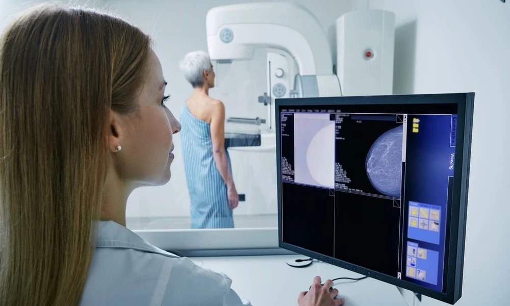
For millions of women worldwide, the annual mammogram represents a crucial checkpoint in breast health management. Yet for a significant portion of the population-those with dense breast tissue, genetic predispositions, or previous breast cancer diagnoses-standard mammography alone may not provide adequate screening. Understanding when additional imaging becomes necessary, and what options exist beyond conventional X-ray technology, can make the difference between early detection and a missed diagnosis.
The Limitations of Standard Mammography
Mammography has been the cornerstone of breast cancer screening for decades, and it remains highly effective for many women. The technology uses low-dose X-rays to detect abnormalities in breast tissue, including calcifications and masses that may indicate cancer. However, mammography faces a significant challenge when imaging dense breast tissue. Dense breasts contain more glandular and fibrous tissue relative to fatty tissue, and both dense tissue and tumors appear white on a mammogram, making it difficult to distinguish between normal tissue and potential abnormalities.
Research indicates that approximately 40 to 50 percent of women have dense breast tissue, though many are unaware of their breast density until they receive their mammogram results. Women with extremely dense breasts face both a higher risk of developing breast cancer and a greater likelihood that cancers will be masked on standard mammograms. This dual challenge has prompted medical professionals to recommend supplemental screening methods for high-risk populations.
Who Needs Enhanced Breast Imaging
Several factors may indicate that a woman should consider imaging beyond standard mammography. Women with heterogeneously dense or extremely dense breast tissue typically receive notification of their density category following a mammogram, and this information should prompt a conversation with their healthcare provider about supplemental screening options. Those with a personal history of breast cancer, particularly if diagnosed at a young age, often require more sensitive imaging to monitor for recurrence.
Genetic factors also play a crucial role in screening decisions. Women who carry BRCA1 or BRCA2 mutations face substantially elevated breast cancer risk-up to 72 percent lifetime risk for BRCA1 carriers and up to 69 percent for BRCA2 carriers. Other genetic mutations, including those in the PALB2, TP53, and PTEN genes, similarly warrant enhanced surveillance. Family history matters even without identified genetic mutations; women with multiple first-degree relatives diagnosed with breast or ovarian cancer should discuss intensive screening protocols with their physicians.
Advanced Imaging Options for High-Risk Screening
When standard mammography proves insufficient, several advanced imaging modalities can provide clearer visualization of breast tissue. Magnetic resonance imaging has emerged as a particularly valuable tool for high-risk screening, offering superior sensitivity compared to mammography alone. Unlike mammography, which uses X-rays, this technology employs powerful magnets and radio waves to create detailed cross-sectional images of breast tissue. The contrast between different tissue types allows radiologists to identify small lesions that might be invisible on conventional imaging.
For women in Abu Dhabi seeking comprehensive breast health evaluation, Cleveland Clinic Abu Dhabi offers diagnostic services including the mri breast scan, which provides detailed imaging for patients with dense breast tissue, genetic predispositions, or other risk factors requiring enhanced surveillance. This multi-specialty hospital delivers cancer care and diagnostic imaging services to patients throughout the UAE, combining advanced technology with specialized expertise in breast health management.
Ultrasound represents another supplemental option, particularly useful for characterizing masses identified on mammography or for screening women with dense breasts. This technology uses sound waves to create images of breast tissue and can distinguish between solid masses and fluid-filled cysts. Automated whole-breast ultrasound systems have improved the efficiency and consistency of ultrasound screening, though interpretation still requires specialized training.
The Science Behind Enhanced Detection
Recent research has validated the clinical value of supplemental screening for appropriate populations. A comprehensive analysis published in The Lancet Regional Health 00017-8/fulltext) examined screening approaches across diverse populations, highlighting the importance of tailoring imaging strategies to individual risk profiles. The evidence demonstrates that combined screening approaches detect significantly more cancers in high-risk women compared to mammography alone, though at the cost of increased false positives and additional imaging procedures.
The trade-off between sensitivity and specificity remains central to screening decisions. More sensitive imaging modalities identify more cancers, including very small or early-stage tumors, but they also flag more benign findings that require follow-up. This can lead to additional biopsies and procedures, along with the anxiety that accompanies uncertain results. Medical professionals must balance the benefit of early cancer detection against the potential harms of overdiagnosis and unnecessary interventions.
Making Informed Screening Decisions
Choosing the appropriate screening strategy requires understanding your personal risk profile and discussing options with qualified healthcare providers. Begin by gathering information about your family history, including any relatives diagnosed with breast, ovarian, or related cancers, along with their ages at diagnosis. If your family history suggests hereditary cancer risk, genetic counseling can help determine whether testing for known mutations is appropriate.
When you receive mammogram results indicating dense breast tissue, ask your radiologist or primary care physician about supplemental screening recommendations specific to your situation. Not all women with dense breasts require additional imaging, particularly if they have no other risk factors. The decision depends on the degree of density, personal and family history, and other individual risk factors.
Financial and Insurance Considerations
Healthcare coverage for supplemental breast imaging varies significantly depending on location, insurance provider, and specific risk factors. Many insurance plans cover enhanced screening for women with documented high risk, such as genetic mutations or strong family histories, but coverage for supplemental imaging based solely on breast density remains inconsistent. Understanding your insurance benefits before scheduling additional imaging can prevent unexpected costs. Resources like reviews of health plan providers can help you evaluate coverage options and identify plans that support comprehensive breast health screening.
Some facilities offer cash-pay options or payment plans for patients whose insurance does not cover supplemental screening, though costs can range widely depending on the imaging type and facility. When considering out-of-pocket screening, weigh the financial investment against your personal risk profile and the potential benefit of earlier detection.
Creating Your Personal Screening Plan
An effective breast health strategy extends beyond periodic imaging. Monthly self-examinations help you become familiar with the normal texture and appearance of your breasts, making it easier to notice changes between screening appointments. While self-exams alone cannot replace professional screening, they provide an additional layer of surveillance that can prompt earlier medical evaluation when changes occur.
Maintain consistent communication with your healthcare team about any breast changes, regardless of how minor they may seem. New lumps, skin changes, nipple discharge, or persistent pain warrant evaluation even if your most recent mammogram was normal. Interval cancers-those that develop between scheduled screening appointments-do occur, and prompt attention to symptoms can facilitate earlier diagnosis.
For women with significantly elevated risk, some physicians recommend alternating mammography with other imaging modalities at six-month intervals rather than annual screening alone. This staggered approach provides more frequent surveillance while utilizing different technologies that may detect changes missed by the other modality. The specific schedule depends on individual risk factors and should be developed in consultation with breast health specialists.
Moving Forward with Confidence
Breast cancer screening decisions have become increasingly personalized as our understanding of risk factors and imaging capabilities has evolved. While the expanding array of options can feel overwhelming, this sophistication allows for better-targeted surveillance that matches individual needs. Women with average risk can continue with standard mammography screening according to recommended guidelines, while those with elevated risk can access enhanced imaging that improves their chances of early detection.
The key lies in active participation in your breast health management. Understand your risk factors, ask questions about your screening options, and work with healthcare providers who take time to explain recommendations and address concerns. Early detection remains the most powerful tool we have against breast cancer, and choosing the right screening approach for your situation maximizes the effectiveness of that tool.

Comments are closed.